Laboratory work No. 1
Study of the microscopic structure of cells and tissues.
Target: familiarization with the structural features, properties and functions of tissues.
Equipment: microscope, ready-made microslides of epithelial, connective, muscle and nervous tissues.
Work progress.
Examine the structure of an animal cell under a microscope.
Consider ready-made tissue microslides.
Presentation of results:
sketch the tissue preparations examined;
fill out the table
| Fabric group | Types of fabrics | Tissue structure | Location | |
Do it conclusion about the structural features of tissues.
Laboratory work № 2
Self-observation of the blink reflex
and the conditions for its manifestation and inhibition.
Target: acquaintance with the structure of the reflex arc of the blink reflex.
Work progress.
Gently touch the inner corner of the eye several times. Determine how many touches the blink reflex will slow down.
Analyze these phenomena and indicate their possible causes. Find out what processes could occur at the synapses of the reflex arc in the first and second cases.
Check the possibility of using volitional effort to slow down the blink reflex. Explain why this was successful.
Remember how the blink reflex manifests itself when a speck gets into the eye. Analyze your behavior from the point of view of the doctrine of forward and backward connections.
Presentation of results:
Using Figure 17, sketch the reflex arc of the blink reflex and indicate its parts.
Do conclusion about the meaning of the blink reflex.
Laboratory work№ 3
Microscopic structure of bone.
Purpose: Study of the microscopic structure of bone.
Equipment : microscope, permanent preparation “Bone tissue”.
Work progress.
Examine bone tissue at low magnification using a microscope. Using Figure 19, A and B, determine: are you considering a transverse or longitudinal section?
Find the tubules through which the vessels and nerves passed. In cross section they look like a transparent circle or oval.
Look for bone cells that are located between the rings and look like black spiders. They secrete plates of bone substance, which are then saturated with mineral salts.
Think about why a compact substance consists of numerous tubes with strong walls. How does this contribute to bone strength with the least amount of material and bone mass required? Why is the airframe made from durable duralumin tubular structures, and not from sheet metal?
Presentation of results:
Draw a longitudinal and transverse section of the microscopic structure of the bone.
Do conclusion
Laboratory work№ 4
Muscles of the human body.
Goal: acquaintance with the structure of the muscles of the human body.
Equipment: tables, drawings, textbook.
Work progress.
Using drawings and anatomical descriptions, identify the location of muscle groups and the movements they perform.
I. Head muscles(according to Figure 35).
Mimic muscles attach to bones, skin, or just To skin, chewable– to the bones of the fixed part of the skull and to the lower jaw.
Task 1. Determine the function of the temporal muscles. Place your hands on your temples and make chewing movements. The muscle tenses as it lifts the lower jaw upward. Find the masseter muscle. It is located near the jaw joints, about 1 cm in front of them. Determine: are the temporal and masticatory muscles synergists or antagonists?
Task 2. Get to know the function of facial muscles. Take a mirror and wrinkle your forehead, which is what we do when we are unhappy or when we are thinking. Reduced supracranial muscle. Find it in the picture. Observe the function orbicularis oculi muscle And orbicularis oris muscle. The first one closes the eye, the second one closes the mouth.
II. Sternocleidomastoid muscle on the front surface of the neck (according to Figure 35).
Task 3. Turn your head to the right and feel the left sternocleidomastoid muscle. Turn your head to the left and find the right one. These muscles turn the head left and right, acting as antagonists, but when contracted together, they become synergists and lower the head down.
III. Muscles front of the torso (according to Figure 36).
Task 4. Find pectoralis major muscle. This paired muscle tenses if you bend your arms at the elbow and forcefully fold them on your chest.
Task 5. Consider in the figure the abdominal muscles that form abdominal press They are involved in breathing, bending the body to the sides and forward, and transferring the body from a lying to a sitting position with fixed legs.
Task 6. Find intercostal muscles: external ones inhale, internal ones exhale.
IV. Muscles back of the body (according to Figure 36).
Task 7. Find in the picture trapezius muscle. If you squeeze your shoulder blades and throw your head back, it will be tense.
Task 8. Find latissimus dorsi muscle. She lowers her shoulders and moves her arms behind her back.
Task 9. Along the spine are deep back muscles. They straighten the body, tilting the body back. Determine their position.
Exercise10. Find gluteal muscles. They abduct the hip. The deep muscles of the back and gluteal muscles in humans are most strongly developed due to upright posture. They resist gravity.
V Muscles hands (according to figures 28, 34 and 36).
Exercise 11. Find in the picture deltoid muscle. It is located above the shoulder joint and moves the arm to the side to a horizontal position.
Exercise 12. Find double-headed And triceps shoulder muscles. Are they antagonists or synergists?
Exercise13. Forearm muscles. To understand their function, place your hand on a table, palm side down. Press it to the table or clench your hand into a fist and unclench it. You will feel the muscles in your forearm contract. This happens because there are muscles located on the side of the palm of the forearm, flexing the hand and fingers, A straightening them are located on the back of the forearm.
Task 14. Feel near the wrist joint from the palmar surface of the tendons that go to the muscles of the fingers. Think about why these muscles are on the forearm and not on the hand.
VI. Leg muscles (according to Figure 36).
Task 15. On the front surface of the thigh there is a very powerful quadriceps femoris muscle. Find it in the picture. She bends her leg at the hip joint and extends it at the knee. To imagine its function, you need to imagine a football player hitting the ball. Its antagonist is the gluteal muscles. They move their leg back. Acting as synergists, both of these muscles hold the body in an upright position, fixing the hip joints.
There are three muscles on the back of the thigh that flex the leg at the knee.
Task 16. Stand up on your toes, you feel tense calf muscles. They are located on the back of the lower leg. These muscles are well developed because they support the body in an upright position and are involved in walking, running, and jumping.
Presentation of results:
Label the muscles in the picture.
Draw a conclusion.
Laboratory work№ 5
Fatigue during static and dynamic work.
Purpose: observation and identification of signs of fatigue during static work.
Equipment : stopwatch, weight 4-5 kg (if you take a briefcase with books, you must first determine its mass).
Work progress.
The subject stands facing the class, extends his arm to the side strictly horizontally. Chalk on the board marks the level at which the hand is located. After preparations, the stopwatch starts on command, and the subject begins to hold the load at the level of the mark. The starting time is indicated in the first line of the table. Then the phases of fatigue are determined and their time is also indicated. It turns out how long it takes for extreme fatigue to occur. This indicator is recorded.
Find out how long it takes to become extremely tired.
Presentation of results:
Write the results in the table
| Static work | Signs of fatigue | |
| No fatigue | The hand with the load is motionless | |
| First phase of fatigue | The hand drops, then jerks back to its original place. | |
| Second phase of fatigue | Hand trembling, loss of coordination, body swaying, facial flushing, sweating | |
| Extreme fatigue | The hand with the load goes down; experience ends |
Draw a conclusion:
Explain the difference between dynamic and static work.
Laboratory work№ 6
Detection of postural disorders.
Goal: to identify postural disorders.
Equipment : measuring tape.
Work progress.
To identify a stoop (round back), use a measuring tape to measure the distance between the most distant points of the left and right shoulder, 3-5 cm down from the shoulder joint, from the chest And from the back. Divide the first result by the second. If the result is a number close to or greater than one, then there are no violations. Getting a number less than one indicates poor posture.
Stand with your back to the wall so that your heels, shins, pelvis and shoulder blades touch the wall. Try sticking your fist between the wall and your lower back. If it goes away, there is a violation of posture. If only the palm passes through, the posture is normal.
Draw a conclusion.
L laboratory work № 7
Detection of flat feet
(work is done at home).
Target: identify flat feet.
Equipment: a bowl of water, a sheet of paper, a felt-tip pen or a simple
pencil.
Movework.
Stand on a piece of paper with your wet foot. Trace the contours of the trace with a felt-tip pen or a simple pencil.
Find the center of the heel and the center of the third toe. Connect the two points found with a straight line. If in the narrow part the footprint does not go beyond the line, there is no flat foot (Fig. 39).
Laboratory work№ 8
Examination of human and frog blood under a microscope.
Goal: familiarization with the structural features of frog and human blood.
Equipment: ready-made microspecimen of “Frog Blood”, temporary microspecimen of human blood, microscope.
Work progress.
Consider the microslide “Frog Blood”.
Find the red blood cells, pay attention to their size and shape.
Examine a microscopic specimen of human blood.
Find red blood cells, pay attention to their color and shape.
Presentation of results:
Compare frog and human red blood cells and record the results in a table.
| Erythrocyte | Cell diameter, µm | Cell shape | Presence of a kernel | Cytoplasmic staining |
| Human | ||||
Draw a conclusion: Why does human blood carry more oxygen per unit time than frog blood?
Laboratory work№ 9
Position of venous valves in the lowered and raised arm. Changes in tissues due to constrictions that impede blood circulation.
Purpose: familiarization with the position of venous valves in the lowered and raised arm, with changes in tissues due to constrictions that impede blood circulation.
Equipment: pharmaceutical rubber ring or thread.
Work progress.
I. Function of venous valves.
Preliminary clarifications. If the arm is lowered, the venous valves prevent blood from flowing down. The valves open only after enough blood has accumulated in the underlying segments to open the venous valve and allow blood to flow upward into the next segment. Therefore, the veins through which blood moves against gravity are always swollen.
Raise one hand up and the other down. After a minute, place both hands on the table.
Why did the raised hand turn pale, and the lowered hand turn red? Were the vein valves closed in the raised or lowered arm?
II. Changes in tissues due to constrictions that impede blood circulation (according to Figure 52).
Preliminary clarifications.Constriction of a limb makes it difficult
outflow of blood through the veins and lymph through the lymphatic vessels. Dilation of blood capillaries and veins leads to redness,
and then to blueness of the part of the organ isolated by the constriction.
Subsequently, this part of the organ becomes white due to the release
blood plasma into the intercellular spaces, since the pressure
blood increases (since there is no outflow of blood), and the outflow of lymph
lymphatic vessels are also blocked. Tissue fluid
accumulates, squeezing cells. The organ becomes dense
touch. The beginning of oxygen starvation of tissues is subjectively felt as “crawling goosebumps” or tingling. The functioning of the receptors is disrupted.
Screw a rubber ring onto your finger or tie your finger with a thread. Notice the change in color of the finger. Why does it turn red first, then purple, and then white? Why are there signs of oxygen deficiency? How do they manifest themselves? Touch an object with your overstretched finger. The finger seems somehow cottony. Why is sensitivity impaired? Why are the tissues of the finger thickened? Remove the bandage and massage your finger towards the heart. What does this technique achieve?
Draw a conclusion by answering the question:
Why is it harmful to tighten your belt and wear tight shoes?
Laboratory work No. 10
Determination of blood flow speed in the vessels of the nail bed.
Goal: learn to determine the speed of blood flow in the vessels of the nail bed.
Equipment: stopwatch, centimeter ruler.
Preliminary clarifications. The vessels of the nail bed include not only capillaries, but also tiny arteries called arterioles. To determine the speed of blood flow in these vessels, you need to find out the path length - S, which blood will travel from the root of the nail to its top, and the time - t, which she will need for this. Then according to the formula V=S
we will be able to find out the average speed of blood flow in the vessels of the nail bed.
Work progress.
Let's measure the length of the nail from the base to the top, excluding the transparent part of the nail, which is usually cut off: there are no vessels under it.
Let's determine the time it takes for blood to cover the total distance. To do this, use your index finger to press the nail plate of your thumb so that it turns white. In this case, the blood will be forced out of the vessels of the nail bed. Now let’s release the compressed nail and measure the time it takes for it to turn red. This moment will tell us the time during which the blood has traveled its way.
Presentation of results:
calculate the blood flow speed using the formula.
Draw a conclusion:
Compare the obtained data with the speed of blood flow in the aorta. Explain the difference.
Evaluation of results
Most people get about 1-0.5 cm/s. This is 50-100 times less than in the aorta, and 25-50 times less than in the vena cava. The slow flow of blood in the capillaries allows tissues to receive nutrients and oxygen from the blood and give it carbon dioxide and waste products.
Laboratory work№ 11
Functional test: reaction of the cardiovascular system to dosed load.
Purpose: determining the dependence of heart rate on physical activity.
Preliminary clarifications. To do this, measure the heart rate (HR) at rest and after dosed exercise. Based on a large statistical material, it was found that in healthy adolescents (after 20 squats), heart rate increases by "/ 3 compared to the resting state and normalizes 2-3 minutes after the end of work. Knowing these data, you can check the state of your cardiovascular system.
Work progress.
Measure your resting heart rate. To do this, take 3-4 measurements per
10 s and multiply the average value by 6. Record the result.
Do 20 squats at a fast pace, sit down and immediately measure your heart rate for 10 seconds after the load. Then after 30 s, 60 s, 90, 120. 150, 180 s. Enter all results into a table.
| Pulse immediately after work | Pulse at intervals, s |
||||||
Based on the data obtained, construct a graph; Plot time on the abscissa axis, and heart rate on the ordinate axis.
Evaluation of results. The results are good if the heart rate after squats increased by 1/3 or less than the resting results; if half, the results are average, and if more than half, the results are unsatisfactory.
Laboratory work No. 12
Measurement of chest circumference during inhalation and exhalation.
Purpose: measurement of chest circumference.
Equipment: measuring tape.
Work progress.
The subject is asked to raise his arms and a measuring tape is placed so that on the back it touches the corners of the shoulder blades, and on the chest it passes along the lower edge of the nipple circles in men and above the mammary glands in women. During the measurement, your arms should be lowered.
Inhalation measurement. Take a deep breath. You can’t strain your muscles and don’t raise your shoulders.
Exhalation measurement. Take a deep breath. Don't droop your shoulders, don't slouch.
Presentation of results:
Enter the obtained data into the table.
Calculate the difference in chest circumference.
| Inhalation measurement. | Exhalation measurement. | |
Normally, the difference in chest circumference in a state of deep inspiration and in a state of deep exhalation in adults is 6-9 cm.
Laboratory work No. 13
The action of salivary enzymes on starch.
Target: show the ability of saliva to digest carbohydrates.
Equipment: starched bandage, cut into pieces 10 cm long, cotton wool, matches, saucer, pharmaceutical iodine (5%), water.
Preliminary clarifications. The purpose of this experiment is to show that salivary enzymes are capable of breaking down starch. It is known that starch with iodine gives an intense blue color, by which it is easy to find out where it was preserved. When starch is treated with salivary enzymes, it is destroyed if the enzymes are active. No starch remains in these places, so they are not stained with iodine and remain light.
Work progress.
Prepare a reagent for starch - iodine water. To this end, pour water into a saucer and add a few drops of iodine (pharmacy 5% alcohol solution) until you obtain a liquid the color of strongly brewed tea.
Wrap cotton wool around a match, moisten it with saliva, and then use this cotton wool and saliva to write a letter on a starched bandage.
Hold the straightened bandage in your hands and hold it for a while until it warms up (1-2 minutes).
Dip the bandage into iodine water, straightening it thoroughly. The areas where starch remains will turn blue, and the areas treated with saliva will remain white, since the starch in them has broken down into glucose, which, under the influence of iodine, does not give a blue color.
If the experiment was successful, you will get a white letter on a blue background.
Draw a conclusion by answering the questions:
What was the substrate and what was the enzyme when you wrote the letters on the bandage?
Could this experiment produce a blue letter on a white background?
Will saliva break down starch if it is boiled?
Laboratory work No.14
Establishment of the relationship between the load and the level of energy metabolism based on the results of a functional test with breath holding before and after the load.
Target: establish the relationship between the load and the level of energy metabolism.
Equipment:
Preliminary remarks. It is known that the intensity of respiration is affected by decay products, in particular carbon dioxide, which is formed as a result of biological oxidation. It has a humoral effect on the respiratory center. When you hold your breath, metabolism in the tissues does not stop, and carbon dioxide continues to be released. When its concentration in the blood reaches a certain critical level, involuntary restoration of breathing occurs. If you hold your breath after work, for example, after 20 squats, it will recover faster, because during squats, biological oxidation occurs more intensely, and more carbon dioxide accumulates by the beginning of the second breath hold.
However, for trained people the difference between these results will be smaller than for untrained people. One of the reasons is that in untrained people, usually, along with the muscles that provide the desired movement, many other muscles that are not related to it contract. Sticky movements are inhibited during training due to improved regulation by the nervous system. Thus, this functional test shows not only the state of a person’s respiratory and cardiovascular systems, but also the degree of his fitness.
Experiment protocol(time is measured in seconds)
Breath holding time at rest (A).
Breath holding time after 20 squats (B).
The percentage of the second result to the first B / A X 100%.
Time for holding your breath and restoring your breath after a minute's rest (C).
Percentage ratio of the third result to the first with / A x 100%.
Work progress.
While sitting, hold your breath as you inhale for as long as possible. Start the stopwatch (deep breathing before the experiment is not allowed!).
Turn off the stopwatch as soon as you regain your breath. Write down the result. Rest 5 minutes.
Stand up and do 20 squats in 30 seconds.
Inhale, quickly hold your breath and start the stopwatch, without waiting for your breathing to calm down, sit on a chair.
Turn off the stopwatch when breathing returns. Write down the result.
After a minute, repeat the first test. Write down the result.
Make calculations in your notebook using the formulas given in paragraphs 3 and 5 of the protocol. Compare your results with the table and determine which category you would place yourself in.
Results of a functional test with breath holding before and after exercise for categories of subjects with different degrees of training.
| Holding your breath |
|||
| A - at rest | B – after work | C – after rest |
|
| V/A X 100%. | s/A x 100%. |
||
| Healthy trained | More than 50% of the first result | More than 100% of the first result |
|
| Healthy untrained | 30-50% of the first result | 70-100% of the first result |
|
| With health problems | Less than 30% of the first result | Less than 70% of the first result |
|
Draw a conclusion by answering the questions:
Why does carbon dioxide accumulate in the blood when you hold your breath?
How does carbon dioxide affect the respiratory center?
Why are these effects called humoral?
Why is it possible to hold your breath for less time after work than at rest?
Why does energy metabolism occur more economically in a trained person than in an untrained person?
Laboratory work No. 15
Preparation of food rations depending on energy expenditure.
Goal: learn competently, create a daily food ration for teenagers.
Equipment: tables of the chemical composition of food products and calorie content, energy needs of children and adolescents of various ages, daily norms of proteins, fats and carbohydrates in the food of children and adolescents.
Work progress.
Make a daily food ration for teenagers 15-16 years old.
Write the calculation results in the table.
(Work is organized in groups. 1-2 – breakfasts, 3 – lunch, 4 – dinner)
Composition of the daily diet.
| Diet | Name of the dish | Products needed for its preparation | Calorie content, kJ |
|||||
| 1st breakfast | ||||||||
| 2nd breakfast | ||||||||
Tables.
Daily energy requirement of children and adolescents of various ages (J)
| Age, years | Total based on average body weight |
| 6720000 - 7560000 |
|
| 7560000 - 9660000 |
|
| 9450000 - 12180000 |
|
| 11760000 - 13860000 |
|
| 13440000 - 14700000 |
Daily norms of proteins, fats and carbohydrates in the food of children and adolescents.
| Age, years | Carbohydrates, g |
||
Composition of food products and their calorie content
| Product name | Carbohydrates | Calorie content per 100 g of product, J |
|||
| in percent |
|||||
| Tangerines | |||||
| Refined sugar | |||||
| Sunflower oil | |||||
| Butter | |||||
| Curd mass | |||||
| Fat cottage cheese | |||||
| Ice cream | |||||
| Beef meat | |||||
| Lamb meat | |||||
| Meat, lean pork | |||||
| Amateur sausage | |||||
| Red caviar | |||||
| Eggplant caviar | |||||
| Buckwheat | |||||
| Semolina | |||||
| Pasta | |||||
| Rye bread | |||||
| Wheat bread | |||||
| Potato | |||||
| Fresh cabbage | |||||
| Sauerkraut | |||||
| Green onion | |||||
| Fresh cucumbers | |||||
| Pickled cucumbers | |||||
| Tomatoes | |||||
| Oranges | |||||
| Grape | |||||
Laboratory work No.16
Finger-nose test and features of movements associated with the functions of the cerebellum and midbrain
Target: Observation of muscle coordination carried out by the cerebellum during the cerebellar finger-nose test.
Work progress.
Close your eyes. Extend the index finger of your right hand forward, which you should hold in front of you. Touch your index finger to the tip of your nose. Change the position of your hand and repeat the experiment. Do the same with your left hand, alternating fingers and hand position. In all cases, the finger hits the target, although the trajectory of movement in each individual case is not the same. With normal functioning of the cerebellum, movements are precise and fast. In persons with a damaged cerebellum, the hand moves in separate jerks, trembles before hitting the target, and misses are frequent.
Answer the questions:
1. What parts does the brain consist of?
What are the functions of the medulla oblongata?
What nerve pathways pass through the pons?
What are the functions of the midbrain?
What is the role of the cerebellum in movement?
Laboratory work No.17
Experiments revealing illusions associated with binocularvision.
Target: identification of illusions associated with binocular vision.
Equipment: a tube rolled from a sheet of paper.
Work progress.
Place one end of the tube to your right eye. Place your left hand on the other end of the tube so that the tube lies between your thumb and index finger. Both eyes are open and should look into the distance. If the images obtained in the right and left eyes fall on the corresponding areas of the cerebral cortex, an illusion will arise - a “hole in the palm”.
Laboratory work№ 18
Developing the skill of mirror writing as an example of the destruction of the old and the formation of a new dynamic stereotype.
Target: develop mirror writing skills.
Working conditions. The experiment can be carried out alone, but it is better if it is carried out in the presence of other people. Then the emotional components associated with the restructuring of the dynamic stereotype appear more clearly.
Work progress
Measure how many seconds it takes to write a word in cursive, such as “Psychology.” On the right side, enter the time spent.
Invite the subject to write the same word in mirror font: from right to left. You must write in such a way that all elements of the letters are turned in the opposite direction. Make 10 attempts, next to each of them on the right side write the time in seconds.
Registration results
Make a graph. On axis X (abscissa) put the serial number of the attempt on the axis Y (ordinate) - the time that the subject spent writing the next word.
Count how many gaps there were between the letters when writing the word in the usual way, how many gaps there were during the first and subsequent attempts to write the word from right to left. Note in what cases emotional reactions occur: laughter, gestures, an attempt to quit work, etc. Name the number of letters in which elements written in the old way occur.
Laboratory work№ 19
Changing the number of oscillations of the image of a truncated pyramid
under different conditions.
Target: determination of the stability of involuntary attention and attention during active work with an object.
Equipment: stopwatch or watch with a second hand.
Preliminary clarifications. Try to imagine a truncated pyramid with its truncated end facing towards you and away from you. When both images are formed, they will begin to replace each other: the pyramid will seem to be facing towards you, then away from you. Every time the image changes, you must write a dashed line in your notebook without looking at it. You can’t take your eyes off the drawing! By the number of vibrations of these images one can judge the stability of attention. Usually the number of fluctuations of attention per minute is measured. To save time, you can measure the number of oscillations in 30 seconds and double the result. Before conducting the experiment, prepare a table. 
Measuring fluctuations in attention under different conditions
| Attention fluctuations | ||
| Involuntary attention (without installation) | ||
| Voluntary attention (with installation, save the created image) | ||
| Voluntary attention with active working with an object | ||
Work progress.
I. Definition of sustainabilityinvoluntary attention.
Look at the drawing without looking up from it for 30 seconds. Every time you change the image, make a stroke in your notebook. Double the number of attention fluctuations in 30 seconds. Enter both values in the appropriate columns of the table.
II. Image retentionarbitrary attention.
Repeat the experiment, following the same methodology, but try to retain the image that has developed for as long as possible. If it does change, you need to maintain the new image for as long as possible. Count the number of vibrations. Record the results in the protocol.
III. Definition of sustainability attention during active work
With object.
Imagine that the drawing represents a room. The small square is its back wall. Think about how to arrange the furniture: sofa, bed, TV, receiver, etc. Do this work for the same 30 seconds. Do not forget to make a touch every time you change the image, and each time return to the original image and continue to “furnish” the room. You need to “arrange” the furniture mentally, without looking up from the drawing. Enter the results obtained in the table in the appropriate columns.
Discussion of results. Typically, the greatest number of fluctuations in attention is observed with involuntary attention.
With voluntary attention with the attitude to hold the established image, the number of oscillations of attention decreases, but following this instruction requires great effort, because both the picture and the attitude remain the same. Therefore, a person has to constantly struggle with the fading of attention. In the third case, many subjects show virtually no fluctuations in attention, although the image of the pyramid remains the same. This is the result of the fact that each subsequent search creates a new situation, causing a discrepancy between what has been done and what remains to be done. This maintains the stability of attention.
Class: 5
Presentation for the lesson


 Back Forward
Attention! Slide previews are for informational purposes only and may not represent all the features of the presentation. If you are interested in this work, please download the full version.
Introduction
An important role in the study of biology at school is played by laboratory work, which contributes to a better assimilation of knowledge and skills of students, contributes to a deeper and more meaningful study of biology, the formation of practical and research skills, the development of creative thinking, the establishment of connections between theoretical knowledge and practical human activities, and facilitates understanding factual material.
An educational experiment has enormous potential for the comprehensive development of students’ personalities. The experiment includes not only the source of knowledge, but also the method of finding it, familiarization with the primary skills of studying natural objects. During the experiment, students gain an understanding of the scientific method of cognition.
Methodical manual “Laboratory workshop. Biology. 5th grade” is intended for organizing research activities of schoolchildren during biology lessons in the 5th grade. The list of laboratory works presented in the methodological manual corresponds to the content of the textbook “Biology” for the 5th grade of general education institutions (authors: I.N. Ponomareva, I.V. Nikolaev, O.A. Kornilova), which opens a line of biology textbooks for primary schools and included in the “Algorithm for Success” system. The textbook does not exactly correspond the paragraphs to the number of hours allocated for their study. Therefore, fewer paragraphs allow the teacher to use the remaining time to conduct laboratory work.
When conducting laboratory work, health-saving technologies, problem-based learning, and development of research skills are used. During practical classes, students develop such universal learning actions as:
- educational – carry out research activities;
- regulatory – check your actions against the goal and, if necessary, correct errors;
- communicative – listen and hear each other, express your thoughts with sufficient completeness and accuracy in accordance with the tasks and conditions of communication.
In the development of practical classes, a problematic question is posed to schoolchildren, the planned results and the necessary equipment are indicated. Each development has instructions for conducting laboratory work. Before performing laboratory work, it is important to familiarize students with the requirements for their execution ( Appendix 1), with safety rules when performing laboratory work ( appendix 2), with rules for making drawings of natural objects ( Appendix 3).
For visual accompaniment of practical classes, an electronic presentation is attached to this manual ( presentation).
Laboratory work No. 1 “Study of the structure of magnifying devices”
Planned results: learn to find parts of a magnifying glass and microscope and name them; follow the rules for working in the office and handling laboratory equipment; use the text and pictures of the textbook to complete laboratory work.
Problematic question: how did people learn about the existence of single-celled organisms in nature?
Topic: “Study of the structure of magnifying devices.”
Goal: study the device and learn how to work with magnifying devices.
Equipment: hand magnifying glass, microscope, watermelon fruit tissue, ready-made microspecimen of camellia leaf.
Work progress
Task 1
1. Examine a hand-held magnifying glass. Find the main parts (Fig. 1). Find out their purpose.

Rice. 1. Structure of a hand-held magnifying glass
2. Examine the flesh of the watermelon with the naked eye.
3. Examine pieces of watermelon pulp under a magnifying glass. What is the structure of watermelon pulp?
Task 2
1. Examine the microscope. Find the main parts (Fig. 2). Find out their purpose. Get acquainted with the rules of working with a microscope (p. 18 of the textbook).

Rice. 2. Structure of the microscope
2. Examine the finished microslide of a camellia leaf under a microscope. Practice the basic steps of using a microscope.
3. Draw a conclusion about the importance of magnifying devices.
Task 3
1. Calculate the total magnification of the microscope. To do this, multiply the numbers indicating the magnification of the eyepiece and objective.
2. Find out how many times the object you are considering can be magnified using a school microscope.
Laboratory work No. 2 “Introduction to plant cells”
Problematic question: “How is the cell of a living organism structured?”
Instruction card for performing laboratory work for students
Topic: “Introduction to plant cells.”
Purpose: to study the structure of a plant cell.
Equipment: microscope, pipette, slide and cover glass, tweezers, dissecting needle, part of an onion, ready-made microslide of a camellia leaf.
Work progress
Task 1
1. Prepare a microslide of onion skin (Fig. 3). In order to prepare a microslide, read the instructions on p. 23 textbooks.

Rice. 3. Preparation of a microslide of onion skin
2. Examine the preparation under a microscope. Find individual cells. Look at the cells at low magnification and then at high magnification.
3. Draw the cells of the onion skin, indicating the main parts of the plant cell in the drawing (Fig. 4).
1. Cell wall
2. Cytoplasm
3. Vacuoles

Rice. 4. Onion skin cells
4. Draw a conclusion about the structure of a plant cell. What parts of the cell were you able to see under the microscope?
Task 2
Compare onion skin cells and camellia leaf cells. Explain the reasons for the differences in the structure of these cells.
Laboratory work No. 3 “Determination of seed composition”
Planned results: learn to distinguish the main parts of a plant cell; follow the rules for handling laboratory equipment; use the text and pictures of the textbook to complete laboratory work.
Problematic question: “How can you find out what substances are part of a cell?”
Instruction card for performing laboratory work for students
Topic: “Determination of seed composition.”
Purpose: to study methods for detecting substances in plant seeds, to study their chemical composition.
Equipment: a glass of water, a pestle, an iodine solution, gauze and paper napkins, a piece of dough, sunflower seeds.
Work progress
Task 1
Find out what organic substances are included in plant seeds using the following instructions (Fig. 5):
1. Place a piece of dough on cheesecloth and make a bag (A). Rinse the dough in a glass of water (B).
2. Open the bag of rinsed dough. Test the dough by touch. The substance that remains on the gauze is gluten or protein.
3. Add 2-3 drops of iodine solution (B) to the cloudy liquid formed in the glass. The liquid turns blue. This proves the presence of starch in it.
4. Place sunflower seeds on a paper towel and crush them using a pestle (D). What appeared on the paper?

Rice. 5. Detection of organic substances in plant seeds
5. Draw a conclusion about what organic substances are included in the seeds.
Task 2
Fill out the table “The importance of organic substances in the cell” using the text “The role of organic substances in the cell” on p. 27 textbooks.
Laboratory work No. 4 “Acquaintance with the external structure of a plant”
Planned results: learn to distinguish and name parts of a flowering plant; sketch a diagram of the structure of a flowering plant; follow the rules for handling laboratory equipment; use the text and pictures of the textbook to complete laboratory work.
Problem question: “What organs does a flowering plant have?”
Instruction card for performing laboratory work for students
Topic: “Acquaintance with the external structure of a plant.”
Purpose: to study the external structure of a flowering plant.
Equipment: hand magnifying glass, herbarium of a flowering plant.
Work progress
Task 1
1. Examine a herbarium specimen of a flowering plant (meadow cornflower). Find the parts of a flowering plant: root, stem, leaves, flowers (Fig. 6).

Rice. 6. Structure of a flowering plant
2. Draw a diagram of the structure of a flowering plant.
3. Draw a conclusion about the structure of a flowering plant. What are the different parts of a flowering plant?
Task 2
Look at the images of horsetail and potatoes (Fig. 7). What organs do these plants have? Why is horsetail classified as a spore plant, and potatoes as a seed plant?

Horsetail Potato
Rice. 7. Representatives of different groups of plants
Laboratory work No. 5 “Observation of the movement of animals”
Planned results: learn to examine single-celled animals under a microscope at low magnification; follow the rules for handling laboratory equipment; use the text and pictures of the textbook to complete laboratory work.
Problematic question: “What is the importance for animals of their ability to move?”
Instruction card for performing laboratory work for students
Topic: “Observing the movement of animals.”
Target: get to know the ways animals move.
Equipment: microscope, slides and coverslips, pipette, cotton wool, glass of water; ciliate culture.
Work progress
Task 1
1. Prepare a microslide with a culture of ciliates (p. 56 of the textbook).
2. Examine the microscopic specimen under a low magnification microscope. Find the ciliates (Fig. 8). Observe their movement. Note the speed and direction of movement.

Rice. 8. Ciliates
Task 2
1. Add a few crystals of table salt to a drop of water with ciliates. Observe how ciliates behave. Explain the behavior of ciliates.
2. Draw a conclusion about the importance of movement for animals.
Literature
- Aleksashina I.Yu. Natural science with basics of ecology: 5th grade: practical. works and their implementation: book. for the teacher / I.Yu. Aleksashina, O.I. Lagutenko, N.I. Oreshchenko. – M.: Education, 2005. – 174 p.: ill. – (Labyrinth).
- Konstantinova I.Yu. Lesson developments in biology. 5th grade. – 2nd ed. – M.: VAKO, 2016. – 128 p. - (To help the school teacher).
- Ponomareva I.N. Biology: 5th grade: methodological manual / I.N. Ponomareva, I.V. Nikolaev, O.A. Kornilov. – M.: Ventana-Graf, 2014. – 80 p.
- Ponomareva I.N. Biology: 5th grade: a textbook for students of general education organizations / I.N. Ponomareva, I.V. Nikolaev, O.A. Kornilov; edited by I.N. Ponomareva. – M.: Ventana-Graf, 2013. – 128 p.: ill.
LABORATORY WORK No. 1
Goals:
Equipment and materials:
Work progress:
LABORATORY WORK No. 1
Topic: Preparation of a temporary microslide. The structure of a plant cell.
Goals:
· learn to make microspecimens on your own;
· become familiar with the structure of a plant cell using a microscope.
Equipment and materials:microscope, dissecting needle, slide and cover glass, filter paper, water, onion bulb scales (juicy).
Work progress:
- Study the sequence of preparation of a temporary microslide.
- Take a glass slide and wipe it with gauze.
3. Use a pipette to place 1-2 drops of water onto the slide.
4. Using a dissecting needle, carefully remove a piece of transparent epidermis from the inner surface of the onion scale. Place it in a drop of water and straighten it with the tip of a needle.
5. Cover the epidermis with a coverslip.
6. Use filter paper on the other side to pull off excess solution.
7. Examine the prepared preparation using a microscope, determining the degree of magnification.
8. Sketch 7-8 cells of the onion scale epidermis. Label the membrane, cytoplasm, nucleus, vacuole with numbers.
9 . Write your conclusion, indicating the functions of the organelles you depicted in the diagram. Answer the question: “Is the nucleus in the center of all cells? Why?".
Laboratory work No. 1
Subject: Consideration of spore-bearing, seed-bearing (gymnosperms and angiosperms) plants: cuckoo flax, fern, Scots pine, shepherd's purse, tomatoes.
Target: Consider the external structure of spore and seed plants.
Equipment: Hand magnifying glass, plant herbarium.
Safety precautions:
Use tools related to laboratory equipment only with the permission of the teacher.
Handle the tool carefully, do not let it fall.
After work, put your workplace in order and hand over the equipment to the teacher.
Work progress:
Task 1. Introduction to spore plants
Examine the plant and fern leaf
Indicate what numbers indicate leaves, rhizomes, spores
_____________________
_____________________
_____________________
Conclude why fern is a spore plant.
_____________________________________________________________________________________________________________________________________________________________________________________________________________________________
Rice. 1. Fern is a higher spore plant.
Task 2. Getting to know a flowering plant
Consider a flowering plant (shepherd's purse).
Find its root, stem, leaf, flower.
_______________________________
_______________________________
_______________________________
_______________________________
_______________________________

Conclude why shepherd's purse, tomato, and Scots pine are seed plants.
____________________________________________________________________________________________________________________________________________________________________________________________________________________________________________________________________________
Reference
Plants differ in origin (wild and cultivated), in life expectancy (annual and perennial), in appearance (life forms), in the complexity of body structure (higher and lower) and in body size. Most of them are green. Due to the presence of chlorophyll, they are all capable of forming organic substances and releasing oxygen in the light. All plants are organisms. Seed and spore plants are members of the plant kingdom. Plants that reproduce by spores are called spore-bearing. Plants that produce seeds are called seed.
Seed plants that produce flowers are called flowering plants.
Laboratory work No. 2
Topic: Introduction to magnifying devices and laboratory instruments .
Target: Study the structure of a magnifying glass and microscope and how to work with them.
Equipment: microscope, magnifying glass.
Work progress:
Quests:
How to find out how many times a microscope magnifies?
Consider the magnifying glass and what parts it has.
Familiarize yourself with the rules for using a magnifying glass.
Examine the microscope, find a tube, an eyepiece and a lens with magnifying glasses, a tripod with a stage and mirror, and screws. Find out the meaning of each part.
Familiarize yourself with the rules for using a microscope in the textbook. Practice the sequence of actions when working with a microscope.
Name the components of a microscope and their significance. Fill out the table:
| Microscope part | Meaning |
| Lens | |
| Adjustment screws | |
| Subject table | |

Draw a general conclusion.
Laboratory work No. 3
Topic: Preparation of microscopic preparations of onion skins a, leaf epidermis.
Target : Study the structure of onion skin cells and leaf epidermis.
Equipment: microscope, dissecting needle, glass slide, onion scales, glass of water, gauze.
Quests:
Prepare the slide by wiping it with gauze. Place 1-2 drops of water on a glass slide.
Using a dissecting needle, remove a small piece of transparent skin. Place a piece of peel in a drop of water and straighten it with the tip of a needle.
Examine the prepared preparation under a microscope. Note which parts of the cell you see.
Preparation of onion skin scale preparation.

Examination of a micropreparation under a microscope.
Start examining the prepared preparation at a magnification of 56 times (lens x8, eyepiece x7). Carefully moving the slide across the stage, find a place on the slide where the cells are best visible.
What are you observing? ______________________________________________________________________________________________________________________________________________________________________________________________________________________________________________________________________________________________________________________________________________________________
Examine the cells under a microscope at 300x magnification (x20 objective, x15 eyepiece). What are you observing? ______________________________________________________________________________________________________________________________________________________________________________________________________________________________________________________________________________________________________________________________________________________________
Conclusion:
During laboratory work we ______________________________________________________________
______________________________________________________________________________________________________________________________________________________________________________________________________________________________________________________________________________
Laboratory work No. 4
Topic: Study of the structure of a plant cell using the example of an elodea leaf and leaf skin.
Target: Study the structure of the leaf cell and leaf skin.
Equipment: microscope, ready-made microscopic specimen of a leaf.
Quests:
Examine the preparation under a microscope.
Find organelles in cells (nucleus, vacuoles, chloroplasts)
Draw 2-3 leaf cells, label the membrane, cytoplasm, nucleus, vacuoles and chloroplasts.

Conclusion:
Laboratory work No. 5
Topic: The structure of seeds of monocotyledonous and dicotyledonous plants.
Introduction to the variety of vegetable seeds.
Target: Study the structure of bean and wheat seeds.
Equipment: dry and swollen wheat and bean seeds, Petri dishes.
Quests:
Examine dry and swollen wheat and bean seeds, compare their sizes and external shape.
Remove the skin from the swollen bean seed (explain why the grain skin is not removed).
Examine the embryo, find the cotyledons, germinal root, stalk, bud.
Draw a bean seed and a grain of wheat, label the parts of the seed.
Draw a conclusion: What are the similarities and differences in the structure of seeds of monocots and dicotyledons?

Conclusion.
Consider the seeds of vegetable crops, pay attention to their color, shape, size. Enter this data into the table.
| Name of vegetable crop | Features of seeds |
|
| Color | ||
| Form | ||
| Size |
Laboratory work No. 6
Topic: Study of the external structure of roots in seedlings (pumpkin peas, beans and wheat.)
Target: Study the structure of the roots of beans and wheat.
Equipment: wheat and bean sprouts, Petri dishes.
Quests:
Consider the root systems of the proposed plants. How are they different?
Based on the structure of the root system, determine which plant belongs to the monocots and which to the dicotyledons.
Fill out the table and draw a conclusion.
| Plant name | Root system type | Features of the structure of the root system |
Laboratory work No. 7
Topic: Determination of the growth zone (extension) at the root.
Target: Determine the growth zone at the plant roots.
Equipment: microscope, microslide “root cap and growth zone”.
Quests:
Examine the preparation under a microscope and find the root cap at the end of the root.
Pay attention to the part of the root above the root cap and division zone. What is this part of the root called?
Draw what you saw under the microscope and make notes.
What is the significance of this zone?

Conclusion:
Laboratory work No. 8
Topic: Modification of roots.
Target: Get acquainted with the modifications of roots in different plants.
Equipment: carrot or beet roots, dahlia root tubers, monstera, banyan, orchid designs.
Quests:
Consider the roots and how they were formed.
How did dahlia root tubers form?
Sketch the root vegetable of a carrot or beet and make inscriptions.
What is the significance of modified roots?

Draw a conclusion.
Laboratory work No. 9
Topic: The structure of vegetative and floral (generative) buds.
Target: Study the structure of the buds of different plants.
Equipment: branches of lilac and poplar with swollen buds, a magnifying glass, a dissecting knife.
Quests:
Consider the shoots of different plants.
Cut the buds and examine them under a magnifying glass. Using the drawing, find scales, rudimentary leaves and flowers, a rudimentary stem, and a growth cone.
Draw a cross-section of the kidneys and label the names of its parts.
What do vegetative and generative buds have in common and how do they differ?

Draw a conclusion about the similarities and differences in the structure of vegetative and generative buds. Make a diagram.
Laboratory work No. 10
Topic: External structure of a leaf. Finding stomata on a leaf.
Purpose of the work : study the external structure of simple and complex leaves
Materials : herbarium specimens of plant leaves, drawings.
Work progress:
1. Look at the plants. Find the parts of the leaf.
2. Examine the veins on the leaf blade. Compare them and note the differences
3. Find simple and compound leaves among them.
4. Fill out the table.
5. Draw a conclusion about the similarities and differences in the structure of simple and complex leaves.
| Plants with simple leaves | Plants with compound leaves | Similarities in leaf structure | Differences in leaf structure |

Laboratory work No. 11
topic: Internal structure of a leaf. Leaf modifications.
Purpose of the work : study the internal structure of leaves, consider leaf modifications.
Materials: herbarium specimens of modified plant leaves.
Work progress :
1. Consider the internal structure of the leaf according to the drawing. Remember the structure and significance of leaf cells.
2. Consider the spines of cactus and barberry, tendrils of peas, aloe leaves and sundews. What significance do they have for the plant?
A very interesting sundew plant.
The leaves of insectivorous plants that live on soils are interesting. A small sundew plant grows in peat bogs. Its leaf blades are covered with hairs that secrete a sticky liquid. The sticky droplets, shiny like dew, attract insects. Insects that land on the leaf become stuck in a sticky liquid. First, the hairs, and then the leaf blade, bend and envelop the victim. When the leaf blade and hairs unfold again, only its integument will remain of the insect. The plant leaf will “digest” and absorb all the living tissues of the insect.


Draw a general conclusion.
Laboratory work No. 12
TOPIC: Examination of growth rings on a cross section (cut) of a tree.
Goals. 1. Study the structure of a tree trunk on a transverse cut.
2. Find out how tree rings are formed.
Equipment: cross-cut of a tree, drawings.
Work progress.
Consider cutting a woody stem. Find the growth rings, count them and determine the age of this stem.
Are the tree rings the same thickness? If not, how can you explain this?
Which growth rings are older: those that are closer to the bark, or those that are closer to the pith? Why?
Can you determine the conditions in which the tree grew?
Make a drawing of the saw cut. Indicate the side that the tree faced north and the side that the tree faced south.


Conclusion:
Laboratory work No. 13
Subject:« Consideration of the structure of the rhizome, tuber and bulb »
Target: get acquainted with modified underground shoots.
Equipment: potato tuber; bulb.
Instruction card.
Examine the base and top of the potato tuber. Find which part has more eyes.
Examine the bulb, find the leaves, buds, and bottom.
Sketch them. Label the drawing.
Make a general conclusion about the work:
How do underground shoots differ from roots?
What functions do underground shoots perform?
Onion bulb

Potato tuber

Laboratory work No. 14
Subject:« Consideration of the structure of a flower »
Target: study the structure of a flower.
Equipment: cherry blossom model, pictures of flowering plants.
Instruction card.
Examine the flower, find the peduncle, receptacle, perianth, stamens and pistil.
Determine which perianth is simple or double.
Consider the structure of the pistil, find its parts.
Examine the structure of the stamen, find the anther and filament.
Draw the parts of the flower and label their names and draw a conclusion.

Laboratory work No. 15
Topic: Comparison of flowers of insect-pollinated and wind-pollinated plants .
Target: compare the characteristics of the flowers of these plants.
Equipment: herbariums, drawings of flowering plants.
Quests:
Fill out the table:
Characteristics of wind-pollinated and insect-pollinated plants.
| Signs | Insect-pollinated plants | Wind-fed |
| 1. Large bright flowers | ||
| 2. Small bright flowers collected in inflorescences | ||
| 3. Availability of nectar | ||
| 4. Small, inconspicuous flowers, often collected in inflorescences | ||
| 5. Presence of aroma | ||
| 6. Pollen is small, light, dry, large quantity | ||
| 7. Large, sticky, rough pollen | ||
| 8. Grow in large clusters, forming thickets | ||
| 9. Plants bloom in the spring before the leaves bloom. |
If the named characteristic is characteristic of a given group of plants, a “+” sign is placed, if not, then “-”
Laboratory work No. 16
Topic: Cuttings of indoor plants.
Target: master the methods of propagating indoor plants from cuttings.
Equipment: a glass of water, scissors, a pot of soil.
Work progress:
Carefully remove a stem cutting with 3-4 leaves from the coleus plant.
Remove the bottom two leaves, make a hole in the soil, and place the cutting in the soil so that the bottom node is hidden by the soil.
Sprinkle the cuttings with soil and water gently.
Draw up a protocol of the experiment, draw a conclusion.
Laboratory work No. 17
Topic: Microscopic and external structure of unicellular and multicellular algae.
Target: study unicellular algae and thallus of filamentous algae.
Equipment: microscope, micropreparations of Volvox and Spirogyra.
Work progress:
Examine the Volvox preparation under a microscope, find two flagella, a shell, a chromatophore, and a nucleus.
Draw a cell and label the names of the parts.
Consider Spirogyra, a filamentous algae. Find cells located one after another in one row. The cells are rectangular in shape, have a clearly defined shell, a nucleus, and a chromatophore in the form of a spiral.
Draw a part of the spirogyra filament and label the names of the cell parts.

1. spirogyra
 2. Volvox cell
Conclusion:
Laboratory work No. 18
Topic: External structure of mosses.
Target: study the structure of moss.
Equipment: herbariums of sphagnum, cuckoo flax.
Work progress:
1.Look at the external structure of the moss, find the stem and leaves.
Indicate the shape, location, size and color of the leaves.
Find the spore capsule at the top of the stem. What is the significance of the dispute?
Compare the structure of moss and algae, what are the similarities and differences.

What is indicated by No. 1,2,3,4.
Draw a conclusion:
Laboratory work No. 19
Topic: Study of the external structure of a fern.
Target: familiarization with the structure of ferns, learning to identify their features
Equipment: herbarium fern leaves with sporangia, herbarium fern rhizomes and adventitious roots; fern leaf (growing in the biology classroom); magnifier and microscope; microslide "Fern Sorus".
Work progress.
1.Look at the fern on a herbarium sheet and note the features of its leaves, stem, rhizome and roots.
2. On the lower surface of the fern leaf, find brown tubercles; they contain sporangia with spores.
3.Look at the "fern sorus" under a microscope
4.Answer the questions:
What is the root system of a fern?
How do leaves grow?
Justify that ferns belong to higher spore plants.
CONCLUSION:
Laboratory work No. 20
Target: study of the appearance of shoots, cones and seeds of conifers.
Equipment: pine shoots, spruce shoots, pine cones, spruce cones.
Work progress
1. Consider the appearance of small branches (shoots) of pine and spruce. Indicate their main differences.
2. Study how the needles of these plants are arranged. Find the shortened side shoots of the pine tree that have needles. How many are there on these shoots?
3. Compare pine and spruce needles, their shape, color, size. Studying the structure of cones and seeds
4.Look at the cones of pine and spruce. Point out their differences.
5. Find traces left by seeds on the scales of the cone.
6.Draw a conclusion: fill out the table.
| Signs | Location on the stem |
||||
Laboratory work No. 21
Topic: Study of the structure of cones and seeds of coniferous plants.
Target: study of the structure of cones and seeds of coniferous plants. Equipment: textbook, table “Characteristics of coniferous trees.”
Work progress
1. Consider the shape of the needles and their location on the stem. Measure the length and pay attention to the color.
2. Using the table “Characteristics of coniferous trees,” determine which tree the branch you are considering belongs to.
Signs of coniferous trees:
The needles are long (up to 5-7 cm), sharp, convex on one side and rounded on the other, sitting 2 together... Scots pine.
The needles are short, hard, sharp, tetrahedral, sit singly, cover the entire branch... Spruce
The needles are light green, soft, sit in bunches like tassels, fall out in the winter... Larch
Consider the shape, size, and color of the cones. Fill out the table.
| Needles | Cone |
||||
| location on a branch | Scale shape | density |
|||
Draw a conclusion.
Laboratory work No. 22
Topic: The structure of the flower and fruit of Cruciferous plants.
Target: study of the structure of the flower and fruit of Brassica plants.
Work progress
1. Consider the structure of the plant given to you.
What type of root system is it?
What kind of stem does the plant have?
What kind of leaves does it have?
How are the leaves located on the stem?
What is the veining of the leaves?
2. Examine the flower.
Which perianth: single or double?
Count the number of sepals.
Examine the sepals, do they grow together?
What is the name of the cup of this flower?
Count the number of petals. Consider the whisk. Do the petals grow together? What is the corolla of such a flower called?
Count the number of stamens. Are all stamens the same size?
Write down what numbers indicate the sepals, petals, stamens, and pistil in the picture.
3. Consider the structure of the fetus.
Measure the width and length of the fruit. If the length of the fruit exceeds its width by 3 or more times, then the fruit is a pod; if the width and length are approximately equal, the fruit is a pod.
Indicate the name of the fruit of this plant.
Write down what numbers indicate the fruit valves, septum, and seed in the picture.

Drawing
1. Write down the numbers of characteristics that representatives of the Cruciferous Family possess.
1. Fruit - berry.
2. Inflorescence - brush.
4. The corolla of the flower consists of 5 free petals.
5. Fruit - bean.
6. The corolla of the flower consists of 4 free petals.
7. Inflorescence – head.
8. A flower has 1 pistil and 6 stamens, of which 2 are short and 4 are long.
9. The fruit is a pod or pod.
10. A flower has 1 pistil and 10 stamens.
_____________________________________________
2. Write down the numbers of plants belonging to the cruciferous family.
| 1. Gulyavnik officinalis | 6. White mustard |
| 2. Wild strawberries | 7. White clover |
| 3. Horseradish | 8. Common cherry |
| 4. Peas | 9. Field jar |
| 5. Chamomile | 10. Common cress |
__________________________________________________
3. Make a table “Plants of the Cruciferous family”
| Gooseberry officinalis Levcoyus jaundice Common cress Field mustard White mustard GulyavnikLezelya Ikotnik gray Common shepherd's purse Yarutka field Wild radish |
In plants of the cruciferous family, the flower has a ................................perianth, the calyx consists of....... ... free sepals, the corolla consists of............ petals, stamens........., pistil............... ....Fruit ………………… or …………………...........
5. Make a table, recording the plants of the Cruciferous family that you know:
| Vegetables | Oilseeds | Decorative | Weeds |
Conclusion:
Laboratory work No. 23
Topic: The structure of the flower and fruit of Rosaceae plants.
Target: study of the structure of the flower and fruit of Rosaceae plants.
Work progress
1. Write down the numbers of characteristics of rosaceous plants.
1. The flower has one pistil and six stamens.
2. The corolla of the flower is fused-petalled, consists of 5 stamens.
3. There are many or one pistils in a flower.
4. The corolla of a flower consists of four free petals.
5. There are many stamens in a flower.
6. The corolla of the flower is separate-petaled, consisting of 5 petals of the same shape.
7. The calyx of a flower consists of 4 free sepals.
8. The calyx of a flower consists of 5 free sepals.
2. Write down the numbers of plants in the Rosaceae family.
| 1. Potentilla goose | 6. Common cherry |
| 2. Chamomile | 7. Black nightshade |
| 3. Field jar | 8. Blood red hawthorn |
| 4. Peas | 9. Rowan |
| 5. Common raspberry | 10. Coltsfoot |
3. Make a table “Plants of the Rosaceae family”
| Wild strawberry Cinquefoil erecta Common cuff Common raspberry Rosehip cinnamon Apple tree Manchurian apple tree Rosehip Kokand |
4. Rewrite the sentences by inserting the missing words.
In plants of the Rosaceae family, the flower has a...... perianth, the calyx consists of......... free sepals, the corolla consists of...... .... free petals, stamens.........., pistils........... or............
5. Distribute the names of plants of the Rosaceae family into groups: a) food, b) ornamental, c) medicinal.
Conclusion:
Laboratory work No. 24
Topic: The structure of the flower and fruit of Solanaceae plants.
Target: study of the structure of the flower and fruit of Solanaceae plants.
Work progress
1. Write down the numbers of characteristics that representatives of the Solanaceae Family possess.
2. The corolla of the flower is fused-petaled and consists of 5 petals.
3. The calyx of a flower consists of 4 free sepals.
6. The fruit is an achene.
7. The calyx of the flower is separate-petaled, consists of 5 sepals.
2. Write down the numbers of plants belonging to the nightshade family.
| 1. Datura common | 9. Wheatgrass |
| 2. Dandelion officinalis | 10. Belladonna belladonna |
| 3. Black henbane | 11. Physalis vulgare |
| 4. Meadow chin | 12. White clover |
| 5. Edible lentils 6. Potatoes 7. Annual sunflower 8. Lupine yellow | 13. Field yarutka 14. Tomato 15. Ordinary cuff 16. Annual pepper |
^
In plants of the nightshade family, the flower has a...... perianth, the calyx consists of......... fused sepals, the corolla consists of...... .... fused petals, stamens.........., pistil........... Fruit ………… or............
^ 4. Distribute the names of plants of the nightshade family into groups: a) food, b) ornamental, c) medicinal.
Conclusion:
Laboratory work No. 25
Topic: The structure of the flower and fruit of legume plants.
Target: study of the structure of the flower and fruit of legume plants.
Work progress
1. Write down the numbers of characteristics that representatives of the Legume Family possess.
1. The corolla of the flower is separate-petaled, consists of 5 petals.
2. The corolla of a flower consists of 5 petals, two of which are fused.
3. The calyx of a flower consists of 4 free sepals.
4. A flower has 1 pistil and 5 stamens.
5. A flower has 1 pistil and 10 stamens.
7. The calyx of a flower consists of 5 fused sepals.
8. Fruit - berry or capsule.
9. Fruit - bean.
10. There are nodules on the roots, which store nitrogen.
2. Write down the numbers of plants belonging to the legume family.
| 1. Datura common | 9. Wheatgrass |
| 2. Dandelion officinalis | 10. Belladonna belladonna |
| 3. Black henbane | 11. Physalis vulgare |
| 4. Sweet clover | 12. White clover |
| 5. Edible lentils 6. Yellow acacia 7. Annual sunflower 8. Lupine yellow | 13. Field yarutka 14. Peas 15. Red clover 16. Annual pepper |
3. Rewrite the sentences by inserting the missing words.
In plants of the legume family, the flower has a...... perianth, the calyx consists of......... fused sepals, the corolla consists of...... .... petals, …….. of which are fused, stamens.........., ……… of which are fused, pistil......... Fruit ………
4. Distribute the names of plants of the legume family into groups: a) food, b) ornamental, c) medicinal, d) forage.
Conclusion:
Laboratory work No. 26
Topic: The structure of the flower and fruit of Asteraceae plants.
Target: determine the structural features of flowers and fruits of plants of the Asteraceae family.
Used educational equipment and materials: collection of dried baskets of sunflowers, aster, collection of seeds of string, dandelion, sunflower.
Tasks to complete
1. Consider the proposed materials, describe the structural features of representatives of the Asteraceae family according to the following plan:
Plant names
Types of leaves, their venation and leaf arrangement,
Types of inflorescence
Sizes of plants, their flowers and seeds
2.Draw different types of Asteraceae flowers, indicate the features of their structure.
3. Describe the structure of flowers, indicating their formulas
4. Determine the type of fruit and make a drawing.
5. Conclusion.
Laboratory work No. 27
Topic: “Structure of the flower and fruit of plants of the Liliaceae family”
1. Write down the numbers of characteristics that representatives of the Liliaceae family possess.
1. The corolla of the flower is separate-petaled, consists of 5 petals.
2. The perianth consists of 6 leaflets.
3. The calyx of a flower consists of 4 free sepals.
4. A flower has 1 pistil and 5 stamens.
5. A flower has 1 pistil and 6 stamens.
6. There are 10 stamens, of which 9 are fused.
7. The perianth is simple, fused-petaled or separate-petaled.
8. Fruit - berry or capsule.
9. Fruit - bean.
10. Intercalary growth of the stem is characteristic.
2. Write down the numbers of plants belonging to the lily family.
| 1. Datura common | 9. May lily of the valley |
| 2. Dandelion officinalis | 10. Belladonna belladonna |
| 3. Onions | 11. Crow's eye |
| 4. Sweet clover | 12. White clover |
| 5. Edible lentils 6. Tulip 8. Lupine yellow | 13. Field yarutka 14. Lily curly 15. Red clover 16. Wheat |
3. Rewrite the sentences by inserting the missing words.
In plants of the lily family, flowers have a ……………… or ………..… perianth consisting of ….… leaflets. In a flower...... stamens and...... pistil. Fruit ……… or ……..
4. Distribute the names of plants of the legume family into groups: a) food, b) ornamental, c) medicinal.
Conclusion.
Laboratory work No. 28
Topic: “Structure of the flower and fruit of plants of the Onion family”
Target: studying the structure of the flower and fruit of plants of the Onion family .
Work progress:
1. Consider a flower of the Onion family. Answer the questions?
2. Sketch it and label all parts of the flower. Write down the formula of the Allium flower._________________________________________________________
3.Name the type of fruit in the onion family.
_______________________________________________________________________
Draw a picture of a fruit from the onion family. Label all parts.
5.Draw a conclusion. What is the significance of plants of the Onion family?
________________________________________________________________________________________________________________________________________________________________________________________________________________________________________________________________________________
Laboratory work No. 29
Topic: “The structure of the flower and fruit of plants of the Cereals family”
Target: studying the structure of flowers and fruits of plants of the Poaceae family .
Work progress:
1. Consider a flower of the Poaceae family. Answer the questions?
A) Which perianth: simple or double?_____________________________________________
B) Count the number of sepals.________________________________________________
C) Examine the sepals, do they grow together?________________________
D) Count the number of petals. Consider the whisk. Do the petals grow together? What is the name of the corolla of such a flower?________________________________________________________________________________
Count the number of stamens. Are all stamens the same size?______________________________________________________________________________
2. Sketch it and label all parts of the flower. Write down the formula of the flower Cereals._________________________________________________________
3. Name the types of fruits of the Cereal family.
4 .Fill out the table:
| Technical | construction | Weed and used in everyday life |
|
5.Draw a conclusion. What is the significance of plants of the Poaceae family?
________________________________________________________________________________________________________________________________________________________________________________________________________________________________________________________________________________________________________________________________________________________________________________________________________________
Laboratory work No. 30
Topic: Consideration of the appearance of Bacillus subtilis.
Work progress:
Prepare a microscopic specimen of Bacillus subtilis and examine it under a microscope. Describe the internal structure of Bacillus subtilis.
____________________________________________________________________________________________________________________________________________________________________________________________________________________________________________________________________________________________________________________________________________
Sketch the cells you see. Label all parts.
From the film covering kefir or pickled cucumber, take a sample with the tip of a dissecting needle and place it in a drop of water with a dye on a glass slide. Mix. Cover with a cover slip and examine under a microscope. Make sure the bacteria come in a variety of shapes. Draw the bacteria seen under the microscope.
Draw a conclusion about the diversity of types and forms of prokaryotic cells. ________________________________________________________________________________________________________________________________________________________________________________________________________________________________________________________________________________________________________________________________
Prove that the cells you see are prokaryotic. Compare the cell of a bacterium and a blue-green algae. What do they have in common and how are they different?______________________________________________________________________________________________
Laboratory work No. 31
Topic: Examination of nodules on the roots of leguminous plants.
Work progress:
Dig out of the ground some well-developed legume plant (peas, beans, vetch, clover, etc.), carefully wash its roots from the soil, and you will see nodules on the roots.
Sketch the pattern of nodules on the roots.
Prepare a micropreparation of nitrogen-fixing bacteria from legume plant nodules. Examine them under a microscope. Describe their internal structure, shape, size________________________________________________________________________________________________________________________________________________________________________________________________________________________________________________________________________________________________________________________________________________________________________________________________________________
Draw a picture of nitrogen-fixing bacteria
Draw a conclusion about the benefits and harms of bacteria.__________________________________________________________________________________________
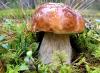
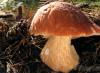
Laboratory work No. 32
Topic: The structure of the fruiting bodies of lamellar and tubular cap mushrooms
Work progress:
Consider the fruiting body of a tubular mushroom. Separate the stump from the cap. Using a dissecting knife, cut the stump lengthwise and use a magnifying glass to examine the internal structure. Sketch the drawing
Let's examine the lower surface of the tubular mushroom cap with a magnifying glass. The holes of the tubes are visible. Special cells called spores are formed in the tubes of the cap. Sketch the drawing.
Let's examine the lower surface of the cap of the agaric mushroom with a magnifying glass. The cap on the underside has plates with spores.
Sketch a drawing of a cap mushroom
Draw a conclusion________________________________________________________________________________________________________________________________________________________________________________________________________________________________________________________________________________________________________________________________
Laboratory work No. 33
Subject: Study of the appearance and microscopic fungus mucor
Work progress:
Examine the mold on the bread with the naked eye. Describe its appearance: note the color of the mold, the smell. Use a dissecting needle to move some of the mold to the side. Note the condition of the food underneath. ________________________________________________________________________________________________________________________________________________________________________________________________________________________________________________________________________________________________________________________________
We are preparing a microscopic specimen of the mycelium of the mucor mushroom. Examine the fungal hyphae, fruiting body and spores under a microscope at 60x magnification. Pay attention to the color of the hyphae and spores. Sketch the drawing.
Prepare a dry (without water) microspecimen of mucor mushroom. Before viewing, place a drop of water under one edge of the cover slip. Watch how the heads of the fungus burst from the water and the spores of the fungus fly away. Sketch the drawing.
Draw a conclusion about the structure of the mucor mushroom.________________________________________________________________________________________________________________________________________________________________________________________________________________________________________________________________________________________________________________________________________________________________________________________________________________________________________________________________________________________________________________________________________________________________________________________________________________________________________________________________________________
Laboratory work No. 34
Topic: Study of the appearance of the fruiting body of the tinder fungus.
Work progress:
1. Examine the appearance of the fruiting body of the mushroom with the naked eye and with a magnifying glass.
2. Examine, sketch and label the parts of the mushroom. Pay attention to the type of spore-bearing layer (tubular or lamellar).
3. Considering that a new layer grows on the fruiting body of the tinder fungus every year, determine its age.________________________________________________________________________________
Describe the structure of the fruiting body of the tinder fungus________________________________________________________________________________________________________________________________________________________________________________________________________________________________________________________________________________________________________________________________________________________________________________
Draw a conclusion about the benefits and harms of the fungus - tinder fungus
__________________________________________________________________
Laboratory work No. 35
“The structure of the thallus of two or three representatives of lichens.”
Target: make sure that lichens are symbiotic organisms based on the study of their structure.
Equipment: magnifying glass, microscope, coverslips and slides, collected lichens of several species.
Work progress:
Try dividing lichens into groups based on appearance.
Explain why you did this?
How are they different from each other?
Using the textbook, look at what groups the authors of the textbook divided lichens into
Prepare a microslide from any lichen. View through a microscope. Compare what you see with the picture in the textbook.

Draw the external internal structure of lichens.
Based on the results of your work, fill out the table:
| Lichen groups | Scale | leafy | bushy |
| Appearance | |||
| Places of settlements | |||
INSTRUCTION No. 2 on labor protection when conducting laboratory work in biology
Students' actions before starting work
Before starting work, the student must: become familiar with the procedure for conducting the experiment and safety measures during work; check the availability and reliability of equipment, utensils, tools, and medications necessary to perform laboratory work.
Safety measures when performing work
When using glass laboratory equipment (test tubes, beakers, slides and cover glasses), strong pressure on the fragile walls of the glassware is not allowed. Slides should be handled lightly by the edges to avoid injury to fingers. When using wet preparations, collections, herbariums, models, stuffed animals, skeletons, as well as various instruments (dynamometers, ergometers, spirometers, microscopes, magnifying glasses and others), handle them carefully and use them only for their intended purpose.
Powdered chemicals used in the experiment must not be handled by hand; special non-metal spoons must be used for this.
When preparing preparations for viewing under a microscope, care must be taken when using piercing and cutting instruments. It is allowed to take tools only by the handles; you cannot point their pointed ends at yourself or your neighbors.
2.4 Liquids remaining after experiments using chemicals must be poured into glass cups or flasks specially prepared for this purpose.
Students' actions after finishing work
Upon completion of work, the student must:
hand over to the teacher or laboratory assistant the instruments and preparations used in laboratory work;
Wash your hands thoroughly with soap.
Laboratory work in biology